Description
- Compact sample compartment design to save lab space
- Uses FTIR detectors – DTGS or MCT
- Available in transmission, reflection and ATR modes
- High throughput optical design
- Simultaneously view and collect spectrum
- Easy-to-use, robust design
- Available for most FTIR spectrometers
- Trinocular with USB camera option
- Low-cost
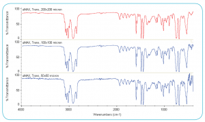
Transmission spectra of polystyrene film at aperture sizes of 200 x 200,
100 x 100, and 50 x 50 microns using the μMAX IR Microscope and the
DTGS detector of the FTIR spectrometer (spectra were collected at 4 cm-1
spectral resolution using a 2-minute collection time).
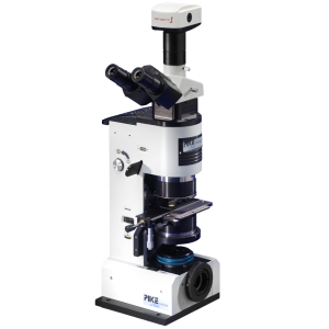
The µMAX™ is an optical design for IR microanalysis providing highperformance sampling at low-cost with exceptional ease-of-use. The µMAX fits into the sample compartment of most FTIR spectrometers. The compact, planar optical layout minimizes the pathlength of the IR beam and thereby maximizes IR throughput.
All operations with the µMAX are intuitive and made even easier with standard Dichroic Optics which provides full viewing of the sample while collecting IR spectra. With Dichroic Optics you can view the sample area and simultaneously search for appropriate IR spectral content – greatly speeding microanalysis. The fully variable X, Y, θ see-through aperture for transmission provides optimized sample dimensioning – for getting the maximum IR signal from every sample.
The µMAX IR microscope uses a 7.45X Schwartzschild objective and condenser to focus the IR beam onto the sample and provide excellent sample visualization – better than 1-micron visible image resolution. An optional CCD camera enables video image projection onto the PC. With the Dichroic Optics of the µMAX and spectral preview of the FTIR software one can view changing IR spectra and sample position in real-time on the PC.
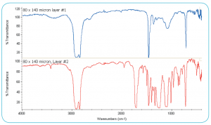
Transmission spectra of polymer laminate sample using DTGS
detector. Samples held in PIKE Micro Compression Cell.
The µMAX is the first sample compartment IR microscope accessory capable of all microsampling modes – transmission, reflection and ATR. The µMAX fits into the sample compartment, using the spectrometers detector for convenience and sampling flexibility. For relatively larger micro samples (100 microns and greater) the DTGS detector provides excellent performance with the µMAX and enables full mid-IR spectral range coverage to 450 cm-1. For smaller micro samples to 20 microns in size an MCT detector is recommended.
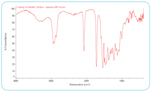
Micro reflection spectrum of a coating on a reflective base metal,
200 x 200 micron sampling area using DTGS detector.
Switching from transmission to reflection on the µMAX is easy with a thumb wheel selection. Reflection sampling area is defined by use of the aperture slide with pre-defined sizes from 40 to 1000 microns. Micro reflection analysis of small areas of interest on reflective surfaces is made easy with the PIKE Technologies µMAX. Simply focus and position the sampling stage, select the sample area with the aperture slide and collect the spectrum. The background spectrum is collected using the same dimension aperture using the gold-surfaced reference slide.
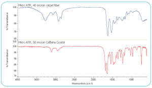
Micro ATR spectra of a 40 micron carpet fiber (upper – blue) and a
50-micron caffeine crystal (lower – red) using DTGS detector
ATR is an excellent sampling option for the µMAX IR microscope. The RotATR™ is a unique, pivot-designed germanium ATR providing easy and precise operation and excellent micro ATR spectra. Focus and select the sample area, rotate the ATR crystal into sample position, make sample contact and collect the IR spectrum. Micro ATR works exceptionally well with the µMAX IR microscope. The 100-micron flat-tipped micro ATR crystal makes intimate contact with the sample easily achieved, providing high spectral quality as seen in the data above.
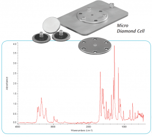
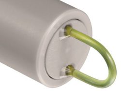
Single drug crystal identified as benzocaine flattened in the Micro
Diamond Cell. Data collected using DTGS detector
The Micro Diamond Cell is an excellent option for use with the µMAX IR Microscope. Tiny chips or fiber segments can be flattened to obtain excellent transmission spectra.